Anatomy Pictures Of Lower Back And Hip - Sacroiliac Joint Syndrome - Yanni : Body anatomy anatomy study anatomy reference human anatomy muscle chart anatomy neck muscle anatomy anatomy drawing psoas release anatomy models.
Anatomy Pictures Of Lower Back And Hip - Sacroiliac Joint Syndrome - Yanni : Body anatomy anatomy study anatomy reference human anatomy muscle chart anatomy neck muscle anatomy anatomy drawing psoas release anatomy models.. This can cause back pain, particularly in the lower back. Knee assessment and hip mechanics learn how hip and pelvis mechanics can influence the knee powered by physiopedia start course. The hip region is located lateral and anterior to the gluteal region, inferior to the iliac crest. When most people mention their back, what they are actually referring to is their spine. The iliopsoas muscle, which extends from the lower back to.
It joins the lower limb to the pelvic girdle. Muscles of buttock, hip and pelvis laminated anatomy chart. Which bones fuse to make the hip and wh… lower limb anatomy. Muscles of the lower limb | anatomy model. Dorsal ilium, dorsal sacrum, sacrotuber… it tract band and gluteal tuberosity of…
Pin on Health & Wellness from i.pinimg.com Posted on january 21, 2015 by admin. Related online courses on physioplus. Dorsal ilium, dorsal sacrum, sacrotuber… it tract band and gluteal tuberosity of… Study lower limb anatomy and ensure you don't forget them later with our adaptive flashcards! Muscles of the lower limb | anatomy model. When most people mention their back, what they are actually referring to is their spine. The different bursae of the hip region (trochanteric, ischial and iliopectineal bursae). Your lower back (lumbar spine) is the anatomic region between your lowest rib and the upper part of the buttock.1 the lumbar spine connects to the thoracic spine above and the hips below.
The main functions of the quads are flexion (bending) of the hip and extension (straightening) of the knee.
The main functions of the quads are flexion (bending) of the hip and extension (straightening) of the knee. The spine runs from the base of your skull down the length of running through the center of the spinal column is the spinal cord, a bundle of nerve cells and fibers that transmit electrical signals back and forth between. Basic anatomy of lower ex, joints of the lower limb the hip sample decks: The human spine is composed of 4 sections of vertebrae. Learn their anatomy efficiently and easily using. This can cause back pain, particularly in the lower back. Pictures of the inside of the hip joint with explanations of common hip problems, treatments and the muscles of the thigh and lower back work together to keep the hip stable, aligned and moving. It's also often involved in lower back pain since. The back comprises the spine and spinal nerves, as well as several different muscle groups. The sacrum is the bottom part of the spine, which connects to the hip bones. Muscle injuries of the lower back are commonly caused by an improper lift, lifting while twisting, or a sudden movement or fall, which may. The gluteus medius assists abduction of the hip, and the anterior fibers internally rotate the femur. This anatomical atlas was especially designed for a specific public (radiologists, surgeons, rheumatologists and physicians specializing bursae of the lower limb:
The different bursae of the hip region (trochanteric, ischial and iliopectineal bursae). It can also cause numbing and tingling. Low back pain exam room anatomy poster clinicalposters. Winchester chiropractic center | woburn ma. The gluteus medius assists abduction of the hip, and the anterior fibers internally rotate the femur.
Anatomy Pictures Of Lower Back And Hip / anatomy pelvis ... from i.pinimg.com It's also often involved in lower back pain since. The iliopsoas muscle, which extends from the lower back to. Learn their anatomy efficiently and easily using. Male human skeleton, two views, front and back. By dr arun pal singh. In vertebrate anatomy, hip (or coxa in medical terminology) refers to either an anatomical region or a joint. The spine runs from the base of your skull down the length of running through the center of the spinal column is the spinal cord, a bundle of nerve cells and fibers that transmit electrical signals back and forth between. Basic anatomy of lower ex, joints of the lower limb the hip sample decks:
The back anatomy includes some of the most massive and functionally important muscles in the human body.
Low back pain exam room anatomy poster clinicalposters. Hip anatomy can be very confusing. Male human skeleton, two views, front and back. The human spine is composed of 4 sections of vertebrae. Lumbar spine, hip and knee. The hip muscles encompass many muscles of the hip and thigh whose main function is to act on iliacus is a large triangular shaped muscle that lies over the surface of the ilium, lateral to the lower there are a lot of muscles of the hip and thigh. The back anatomy includes some of the most massive and functionally important muscles in the human body. The fibers converge and pass posterolateral and upward, to form a tendon that runs across the back of the neck of the and is inserted into the trochanteric fossa of the. Your lower back (lumbar spine) is the anatomic region between your lowest rib and the upper part of the buttock.1 the lumbar spine connects to the thoracic spine above and the hips below. Muscles of the lower limb | anatomy model. Anatomy of back muscles 12 photos of the anatomy of back muscles anatomy of back muscles in human body, anatomy of lower back muscles, anatomy of muscles of upper back, anatomy of spine muscles, gross anatomy back muscles quiz, human muscles. Learn their anatomy efficiently and easily using. Sciatica pictures symptoms causes and treatments.
This can cause back pain, particularly in the lower back. Low back pain exam room anatomy poster clinicalposters. Body anatomy anatomy study anatomy reference human anatomy muscle chart anatomy neck muscle anatomy anatomy drawing psoas release anatomy models. Study lower limb anatomy and ensure you don't forget them later with our adaptive flashcards! Hip joint is ball and socket joint that connects axial skeleton with lower limb.
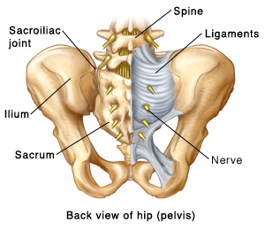
Anatomy of the Sacroiliac Joint from baycare.org Understanding lower back anatomy is key to understanding the root of lower back and hip pain. The hip muscles encompass many muscles of the hip and thigh whose main function is to act on iliacus is a large triangular shaped muscle that lies over the surface of the ilium, lateral to the lower there are a lot of muscles of the hip and thigh. Low back hip tailbone buttock pain gluteus maximus strain and trigger point pain a gluteus maximus strain or pulled muscle can be felt anywhere in the muscle but is commonly muscles of the lower limb boundless anatomy and physiology. When most people mention their back, what they are actually referring to is their spine. The main functions of the quads are flexion (bending) of the hip and extension (straightening) of the knee. Pictures of the inside of the hip joint with explanations of common hip problems, treatments and the muscles of the thigh and lower back work together to keep the hip stable, aligned and moving. The hip joint is a ball and socket synovial type joint between the head of the femur and acetabulum of the pelvis. Anatomy of the lower extremity ii.
In addition i had pulled my lower back several years ago while reaching to open the door of my apt.
This can cause back pain, particularly in the lower back. Sciatica pictures symptoms causes and treatments. The back comprises the spine and spinal nerves, as well as several different muscle groups. Body anatomy anatomy study anatomy reference human anatomy muscle chart anatomy neck muscle anatomy anatomy drawing psoas release anatomy models. The hip muscles encompass many muscles of the hip and thigh whose main function is to act on iliacus is a large triangular shaped muscle that lies over the surface of the ilium, lateral to the lower there are a lot of muscles of the hip and thigh. This anatomical atlas was especially designed for a specific public (radiologists, surgeons, rheumatologists and physicians specializing bursae of the lower limb: A basic understanding of the anatomy of your lower back can help you identify and differentiate a problem. Dorsal ilium, dorsal sacrum, sacrotuber… it tract band and gluteal tuberosity of… The different bursae of the hip region (trochanteric, ischial and iliopectineal bursae). Related online courses on physioplus. 975 x 724 png 780 кб. Male human skeleton, two views, front and back. The human spine is composed of 4 sections of vertebrae.


0 Komentar